Requirements for Imaging
|
|
|
- Shon Merritt
- 6 years ago
- Views:
Transcription
1 Requirements for Imaging Max Seidensticker Universitätsklinikum Magdeburg Klinik für Radiologie & Nuklearmedizin SORAMIC 1
2 Requirements for Imaging SORAMIC: Evaluation of Sorafenib and microtherapy guided by Gd EOB DTPA enhanced MRI in patients with inoperable hepatocellular carcinoma Primary study objective No. 3: To confirm in a 2 step procedure that Primovist enhanced MRI is non inferior (first step) or superior (second step) compared with contrast enhanced multislice CT for stratification of patients to a palliative vs. curative treatment strategy. Secondary study objectives: to compare the number of detected lesions and the diagnostic confidence in Primovist enhanced MRI with contrast enhanced CT to compare Primovist enhanced MRI with contrast enhanced CT regarding the detection of recurrence (patients in the curative study group only) SORAMIC 2
3 Requirements for Imaging Diagnostic substudy Primary endpoint: correct assignment to curative or palliative strategy Secondary endpoint: lesion detection and detection of tumor recurrence in the curative treatment arm SORAMIC 3
4 Imaging: Objectives SORAMIC 4
5 Defining same scanning Ensure Standardization of Imaging data parameters for imaging modalities across sites Consider conflicts with existing local protocols SORAMIC 5
6 Ensure Quality of Imaging data Qualified equipment same equipment for screening & follow up Consistent images over time for a patient (anatomy, modality, parameters) Complete data set Independent evaluation with minimal variability SORAMIC 6
7 Quality assurance: Ensure Quality of Imaging data Validation of CT and MRI scans of the liver before study initiation Imaging work shop, Primovist training Investigator meeting Validation of CT and MRI quality during the study course MRI: Phantom measurements SORAMIC 7
8 Concerning MRI scanners: Ensure Quality of Imaging data large variety of scanners in SORAMIC ( manufacturers / models / age of device ) differences in image quality maybe expected Concerning MRI scanner manufacturers: large variety of SOPs concerning quality assurance (phantoms / sequences) low procedure transparency comparable, transparent and independent quality evaluation has to be established SORAMIC 8

9 Ensure Quality of Imaging data MRI Phantom: ACR MRI Quality Phantom (will be provided) Quality assurance for each MRI Scanner throughout the SORAMIC study. Confirmation of MRI validity in SORAMIC SORAMIC 9
10 Imaging: Screening & Follow Up SORAMIC 10
 IMAGE FOLLOW UP EVERY 2 MONTHS! IMAGE FOLLOW UP NOT MANDATORY!")
11 Screening phase: Primovist enhanced MRI and contrast enhanced CT assessment of disease stage decision on treatment strategy (curative vs. palliative) IMAGE FOLLOW UP EVERY 2 MONTHS! IMAGE FOLLOW UP NOT MANDATORY! SORAMIC 11
 only recurrence to be confirmed by truth panel recurrence: endpoint is reached no recurrence: continued imaging follow up 2 mo 2 mo 2 mo 2 mo 2 mo time SORAMIC 12")
12 Follow Up (curative treatment group): Primovist enhanced MRI and contrast enhanced CT every two months assessment and reading by local investigator (endpoint: Time To Recurrence) only recurrence to be confirmed by truth panel recurrence: endpoint is reached no recurrence: continued imaging follow up 2 mo 2 mo 2 mo 2 mo 2 mo time SORAMIC 12 Recurrence
13 Follow Up (palliative treatment group): Diagnostic imaging is not required in the trial context (endpoint: Overall Survival) Will be performed in investigator s discretion If diagnostic imaging is performed during follow up, results must be reported on the CRF SORAMIC 13
14 HCC Image Characteristics SORAMIC 14
15 Primovist MRI Arterial enhancement plus portal venous washout plus hypointensity in hepatobiliary phase (typical HCC) Arterial enhancement plus portal venous washout with isoto hyperintensity in hepatobiliary phase (well differentiated HCC) Arterial enhancement without portal venous wash out plus hypointensity in hepatobiliary phase (strong indication for HCC) SORAMIC 15
16 Contrast enhanced CT Arterial enhancement plus portal venous washout Mosaic pattern Pseudocapsule (Calcifications, necrosis, hemorrhage, intralesional fat) SORAMIC 16
17 Tumor Recurrence in both modalities Newly detected lesion: longest diameter at least 10mm typical vascular pattern of HCC (arterial enhancement plus portal venous washout) OR Any lesion: with at least 10mm interval growth in subsequent scans Continue follow up scans until verification of tumor recurrence by truth panel! SORAMIC 17
18 Examples SORAMIC 18


19 MRI HCC in cirrhosis 2D T1 w GRE, noncontrast 3D T1 w GRE, FS noncontrast 3D T1 w GRE, FS arterial 3D T1 w GRE, FS portal 3D T1 w GRE, FS late dyn 2D T2 w TSE, FS 3D T1 w GRE, FS 20 min post Primovist SORAMIC 19


20 MRI HCC in cirrhosis 3D T1 w GRE, FS noncontrast 3D T1 w GRE, FS arterial 3D T1 w GRE, FS portal 3D T1 w GRE, FS late dyn 2D T2 w TSE, FS 3D T1 w GRE, FS 20 min post Primovist SORAMIC 20


21 MRI HCC in cirrhosis 3D T1 w GRE, FS noncontrast 3D T1 w GRE, FS arterial 3D T1 w GRE, FS portal 3D T1 w GRE, FS late dyn 2D T2 w TSE, FS 3D T1 w GRE, FS 20 min post Primovist SORAMIC 21

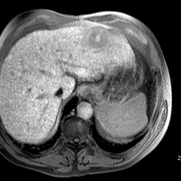
22 CT HCC in cirrhosis MSCT, noncontrast arterial portalvenous SORAMIC 22
23 Requirements: MRI SORAMIC 23
24 Technical Requirements High field MRI (1.5 3T) Phased array surface coil Arms should be positioned overhead, out of field of view (FOV) FOV large enough, just to enclose entire liver (consistent throughout the study) SORAMIC 24
25 Contrast Requirements Contrast Media: Primovist (Gadolinium EOB DTPA) 0.1 ml/kg (10 ml maximum) or mmol/kg via rapid hand or power injector (1,5mL/sec) + 30 ml saline flush (1,5mL/sec) Venous access (preferably 20G) SORAMIC 25

26 Primovist (Gadolinium EOB DTPA) Uptake by hepatocyte and excrete via billiary system Hepatocyte specific CM Combination of dynamic vascular phase and hepatocyte specific late phase imaging Dynamic perfusion information comparable to ECCM Hepatocyte specific phase: improved lesion detection SORAMIC 26
27 Primovist (Gadolinium EOB DTPA) Side effects (<1/100, >1/1000): headache, dizziness, paraesthesia, parosmia, increased blood pressure, flushing, dyspnea, respiratory distress, vomiting, nausea, rash, pruritus, chest pain Electrolyte changes, elevated LFTs Transient QT prolongation Anaphylatic reactions (Nephrogenic systemic fibrosis) All AEs have to be reported to the sponsor SORAMIC 27

28 Standard Protocol Recommendation MRI SORAMIC 28
29 Scanning Parameters: MRI SORAMIC 29
30 General Patient orientation: supine Coil: Phased array coil Scan location / coverage: ensure complete coverage of the liver. Scan FOV: Large (consistent throughout the study), e.g. ±350x350mm Skip /gap (slice spacing): As close to 0% as possible while avoiding cross talk Breath hold: not to exceed 20sec scan time. SORAMIC 30


31 MRI of the liver Start Localizer Should be performed at least in coronal orientation (mandatory) Other orientations are optional SORAMIC 31
32 Scan 1 (Precontrast) T1 w GRE, 2D Slice thickness: 6mm Orientation: Axial Sequence: 2D T1 w gradient echo, breath hold sequence with fat suppression (e.g. WATS, FLASH, SPGR, FFE) Dual (in and opposed) phase imaging optional SORAMIC 32

33 Scan 1 (Precontrast) 2D T1 w gradient echo, breath hold sequence without fat suppression SORAMIC 33
34 Scan 2 (Precontrast) T1 w GRE, 3D Slice thickness: 5mm Orientation: Axial Sequence: 3D T1 w gradient echo, breath hold sequence with fat suppression (e.g. VIBE, THRIVE, LAVA) SORAMIC 34

35 Scan 2 (Precontrast) 3D T1 w gradient echo, breath hold sequence with fat suppression SORAMIC 35
36 Scan 3 (Post-Contrast, Arterial Phase) T1 w GRE, 3D Slice thickness: 5mm Orientation: Axial Sequence: 3D T1 w gradient echo, breath hold sequence with fat suppression (e.g. VIBE, THRIVE, LAVA) Scan Delay Via bolus tracking to ensure arterial phase of the liver (approximately 20 sec. p.i.) SORAMIC 36

37 Scan 3 (Post-Contrast, Arterial Phase) 3D T1 w gradient echo, breath hold technique with fat suppression SORAMIC 37
38 Scan 4 (Post-Contrast, Portal Venous Phase) T1 w GRE, 3D Slice thickness: 5mm Orientation: Axial Sequence: 3D T1 w gradient echo, breath hold sequence with fat suppression (e.g. VIBE, THRIVE, LAVA) Scan Delay Approximately sec. post injection of CM to ensure portal phase of the liver SORAMIC 38
39 Scan 4 (Post-Contrast, Portal Venous Phase) 3D T1 w gradient echo, breath hold technique with fat suppression SORAMIC 39
40 Scan 5 (Post-Contrast, Late Dynamic Phase) T1 w GRE, 3D Slice thickness: 5mm Orientation: Axial Sequence: 3D T1 w gradient echo, breath hold sequence with fat suppression (e.g. VIBE, THRIVE, LAVA) Scan Delay Approximately 120 sec. post injection of CM to ensure equlibrium phase of the liver SORAMIC 40
41 Scan 5 (Post-Contrast, Late Dynamic Phase) 3D T1 w gradient echo, breath hold technique with fat suppression SORAMIC 41
42 Scan 6+7 (Post Contrast) T2 w TSE, 2D Slice thickness: 8mm Orientation: Axial Sequence: 2D T2 w turbo/fast spin echo (TSE, FSE, RARE) respiratory triggered or navigator gated With and without fat suppression SORAMIC 42
,")

43 Scan 6+7 (Post Contrast) 2D T2 w turbo/fast spin echo (TSE), respiratory triggered with fat suppression without fat suppression SORAMIC 43
44 Scan 8 (Hepatobiliary Phase) T1 w GRE, 3D Slice thickness: 6mm Scan delay: at least 20 min post injection Orientation: Coronal Sequence: 3D T1 w gradient echo breath hold sequence With fat suppression (e.g. VIBE, THRIVE, LAVA) SORAMIC 44

45 Scan 8 (Hepatobiliary Phase) 3D T1 w gradient echo, breath hold sequence with fat suppression SORAMIC 45
46 Scan 9 (Hepatobiliary Phase) T1 w GRE, 3D Slice thickness: 5mm Scan delay: at least 20 min post injection Orientation: Axial Sequence: 3D T1 w gradient echo breath hold sequence With fat suppression (e.g. VIBE, THRIVE, LAVA) SORAMIC 46

47 Scan 9 (Hepatobiliary Phase) 3D T1 w gradient echo, breath hold sequence with fat suppression SORAMIC 47
 SORAMIC 48")
48 Scan 10 (Hepatobiliary Phase) T1 w GRE, 2D Slice thickness: 6mm Orientation: Axial Sequence: 2D T1 w gradient echo, breath hold sequence with fat suppression (e.g. WATS, FLASH, SPGR, FFE) SORAMIC 48

49 Scan 10 (Hepatobiliary Phase) 2D T1 w gradient echo, breath hold sequence with fat suppression SORAMIC 49
50 Tips and Tricks: MRI SORAMIC 50
 affecting time of bolus arrival and peak enhancement duration")
51 Enhancement Use bolus detection techniques for proper and individual timing Need trigger delay for best enhancement of lesion, to be defined individually due to individual differences (circulation time, cardiac output) affecting time of bolus arrival and peak enhancement duration SORAMIC 51


52 Dynamic Enhancement arterial portal venous venous Arterial perfusion Portal venous Venous Liver SORAMIC parenchyma 52
")
53 MRI Artifacts To avoid artifacts, arms should be positioned overhead, out of field of view (FOV) SORAMIC 53

54 MRI Artifacts Minimize breathing artifacts Relax and train patient to breath in and breath out Perform exam at breath out Delay between breath out order and start of acquisition Amplitude Respiration Scan Scan Scan SORAMIC 54 t
55 Requirements: CT SORAMIC 55
56 Technical Requirements Helical multislice CT (at least 4 rows) Arms should be positioned overhead, out of field of view (FOV) Scan FOV large Display FOV unique to patient size SORAMIC 56
57 Contrast Media: Contrast Requirements Non ionic agent ( mg/ml Iodide, 300mg/ml recommended) ml + 30 ml saline flush via rapid hand or power injector (at least 3mL/sec) Venous access (preferably 20G) Automatic bolus tracking SORAMIC 57
58 Contrast media Side effects (AE: 3,13%, SAE: 0,004 0,04% (non ionic CM) Katayama H, 1990, Radiology Anaphylactic reaction (pruritus, urticaria, exanthema, erythema, angioedema, flush, dyspnea, hypotension, cardiovascular shock, respiratory arrest) Vasovagal reaction (bradycardia, hypotension, nausea, vomiting) Contrast induced nephropathy Lactic acidosis Extravasation All AEs have to be reported to the sponsor SORAMIC 58
59 General Patient orientation: Supine Scan FOV: Large, complete body diameter (consistent throughout the study) Breathing instructions: One breath hold Time per tube rotation: 1 second or less Acquired slice thickness: 5mm Reconstructed and submitted slice thickness: 5mm Gap (slice spacing): None (i.e. contiguous) Tube voltage (kv):120 Tube current (ma): (anatomically adapted tube current modulation is preferred) Kernel: Use standard abdominal soft tissue kernel SORAMIC 59

60 SORAMIC 60
61 Scanning Parameters: CT SORAMIC 61
62 Scan 1 (Precontrast) Scan coverage: right dome of diaphragma through kidneys (whole liver) SORAMIC 62
")
63 Scan 1 (Precontrast) SORAMIC 63
64 Scan 2 (Postcontrast Arterial Phase) Scan coverage: right dome of diaphragma through kidneys (whole liver) Scan delay: Via bolus tracking to ensure arterial phase of the liver (aortic enhancement between HU) Trigger delay: none (scanning is initiated immediately, <5sec delay for breathing instruction) SORAMIC 64
65 Scan 2 (Postcontrast Arterial Phase) SORAMIC 65
66 Scan 3 (Postcontrast Portal Venous Phase) Scan coverage: right dome of diaphragma through kidneys (whole liver) Scan delay: Start 40 sec after starting injection of contrast media to ensure portal venous phase of the liver SORAMIC 66
")
67 Scan 3 (Postcontrast Portal Venous Phase) SORAMIC 67

68 Scan 4 (Postcontrast Venous Phase) Scan coverage: right dome of diaphragma through kidneys (whole liver) Scan delay: Start 80 sec after starting injection of contrast media to ensure venous phase of the liver SORAMIC 68
69 Scan 4 (Postcontrast Venous Phase) SORAMIC 69
70 Frequent Imaging Issues SORAMIC 70


71 Axial CT images are required SORAMIC 71


72 Field of view should be large enough to view entire anatomy SORAMIC 72

73 Annotations Compromise the unbiased nature of the external review External reviewer should be assessing each patient without any outside influence from site SORAMIC 73

74 Hardcopy films SORAMIC 74
75 Thank you for your attention! SORAMIC 75
RSNA 2006 November 26 to December 1 Chicago. Guest author for ImPACT Dr. Koos Geleijns, Medical Physicist, Leiden University Medical Center.
 RSNA 2006 November 26 to December 1 Chicago Guest author for ImPACT Dr. Koos Geleijns, Medical Physicist, Leiden University Medical Center. Once again, more than 60,000 participants (including professional
2
 1 2 3 4 5 6 7 1. Bauhs, J. A., Vrieze, T. J., Primak, A. N., Bruesewitz, M. R., & McCollough, C. H. (2008). CT Dosimetry: Comparison of Measurement Techniques and Devices1. Radiographics, 28(1), 245-253.
RAD 465 (MRI) Lecture one (Pulse Sequences) Ruba Khushaim MSc
 Lecture one (Pulse Sequences) Ruba Khushaim MSc") RAD 465 (MRI) Lecture one (Pulse Sequences) Ruba Khushaim MSc Outline : Spine echo pulse sequence SE Fast spin echo pulse sequence FSE Inversion recovery pulse sequence IR Gradient pulse sequence GS Pulse
RAD 465 (MRI) Lecture one (Pulse Sequences) Ruba Khushaim MSc Outline : Spine echo pulse sequence SE Fast spin echo pulse sequence FSE Inversion recovery pulse sequence IR Gradient pulse sequence GS Pulse
Request for Proposals
 Request for Proposals Reference: ERDFIAI2012-4006A Caring First Ltd, is a limited liability company providing healthcare services. The company is currently commissioning a private new-build hospital complete
M R I Physics Course. Jerry Allison Ph.D. Chris Wright B.S. Tom Lavin M.S.M.P. Department of Radiology Medical College of Georgia
 M R I Physics Course Jerry Allison Ph.D. Chris Wright B.S. Tom Lavin M.S.M.P. Department of Radiology Medical College of Georgia M R I Physics Course chapter 12 Artifacts and Suppression Techniques Artifacts
Breast MR Imaging and Quality Control
 Breast MR Imaging and Quality Control Donna M. Reeve, MS, DABR, DABMP Department of Imaging Physics Educational Objectives 1. Provide an overview of breast MR imaging and MR-guided biopsy procedures. 2.
MSK Imaging Fundamentals
 MSK Imaging Fundamentals Goals Improve image quality Provide best possible product for our customers Patient Referring clinician Radiologist Reduce number of callback cases Goal reduction of 50% in 3 months
True comfort and flexibility with the power of 3T.
 True comfort and flexibility with the power of 3T. With a large 71 cm aperture and the quietest exams in the industry, the Vantage Titan 3T is the most comfortable 3T MRI system for all of your patients.
Joint ICTP/IAEA Advanced School on Dosimetry in Diagnostic Radiology and its Clinical Implementation May 2009
 2033-17 Joint ICTP/ Advanced School on Dosimetry in Diagnostic Radiology and its Clinical Implementation 11-15 May 2009 Dosimetry for CT 3: Practical Experiences Claire-Louise Chapple Freeman Hospital
Multiparametric MRI Prostate Imaging Protocol November 2015 Full Acquisition Protocol with Parameters GE 3T Magnet with Software Version DV25
 3Plane Loc SSFSE Multiparametric MRI Prostate Imaging Protocol November 2015 Full Acquisition Protocol with Parameters GE 3T Magnet with Software Version DV25 Save Series Scan After acquisition, scroll
Understanding CT image quality
 IAEA RER/9/135 COURSE ON OPTIMIZATION IN COMPUTED TOMOGRAPHY Sofia, Bulgaria, 2017 Understanding CT image quality Dean Pekarovič UMC Ljubljana, Institute of Radiology Quality and Safety office Role of
Image quality in non-gated versus gated reconstruction of tongue motion using Magnetic Resonance Imaging:
 This talk was presented 26 June 2008, at the 22nd International Congress and Exhibition of Computer Assisted Radiology and Surgery, in Barcelona at the Hotel Constanza from June 25 to 28, 2008. See http://kochanski.org/gpk/papers/2008/carstalk.html
PATIENT POSITION IMAGING PARAMETERS
 3 PLANE LOC Patient Entry Feet First Imaging Mode 2D Patient Position Prone Pulse Sequence Gradient Echo Coil Configuration 7breast both MRI Imaging Options Seq, Fast Plane 3-PLANE Acceleration Factor
4/14/2009. The Big Picture of Quality. MRI Quality Assurance and ACR MRI Accreditation Program. Basic Elements for Image Quality.
 The Big Picture of Quality MRI Quality Assurance and ACR MRI Accreditation Program Chen Lin, PhD Indiana University School of Medicine & Clarian Health Partners Diagnosis accuracy Image quality Knowledge
CT Numbers: Think of a number, double it, add 20, divide by 4. Jane Edwards Royal Free Hospital, London
 CT Numbers: Think of a number, double it, add 20, divide by 4 Jane Edwards Royal Free Hospital, London Background 2 different manufacturers CT scanners available on site: GE Lightspeed Plus; Philips Brilliance
Iterative Reconstruction with Philips idose Characterising Image Quality in Attempting to Realise its Potential
 Iterative Reconstruction with Philips idose Characterising Image Quality in Attempting to Realise its Potential Julie Smyth & Philip Doyle Regional Medical Physics Service Outline Preamble Image Quality
Procedure Manual for MRI of the Brain
 Baxter Protocol 161003 SYN RC W H E R E S C I E N C E M E E T S S E R V I C E Baxter Protocol 161003 A Phase 3 Randomized, Double-Blind, Placebo-Controlled Study of the Safety and Effectiveness of Immune
Brilliance CT 64-channel configuration
 Brilliance CT 64-channel configuration with Essence technology The Brilliance CT 64-channel configuration is designed to help you conduct the most advanced multislice CT studies possible. These systems
Phantom Test Guidance for Use of the Small MRI Phantom for the MRI Accreditation Program
 Phantom Test Guidance for Use of the Small MRI Phantom for the MRI Accreditation Program 1 Contents 0.0 INTRODUCTION 4 0.1 Overview and Purpose 4 0.2 The Phantom 4 0.3 The Required Images 5 0.4 The Image
Peacefully quiet. Remarkably fast.
 Peacefully quiet. Remarkably fast. Excellent image quality Streamlined workflow Outstanding patient comfort Canon Medical Systems Vantage Galan 3T offers a transformational experience for you and your
Abstract. Learning Objectives 8/1/2017
 SAM Practical Medical Physics TU-B-201-0 AAPM Annual Meeting 2017 1 Abstract This course will teach the participant to identify common artifacts found clinically in MR, DR, CT, PET, to determine the causes
Scope: All CT staff technologist
 APPROVED BY: Radiology Technical Director Page 1 of 6 Purpose: The QC program assesses relative changes in system performance as determined by the technologist, service engineer, qualified medical physicist,
Peacefully quiet. Remarkably fast.
 Peacefully quiet. Remarkably fast. 2 Excellent image quality Streamlined workflow Outstanding patient comfort Toshiba Medical s Vantage Galan 3T offers a transformational experience for you and your patients
2012 Computed Tomography
 2012 Computed Tomography QUALITY CONTROL MANUAL Radiologist s Section Radiologic Technologist s Section Medical Physicist s Section 2012 Computed Tomography QUALITY CONTROL MANUAL Radiologist s Section
EPI. Thanks to Samantha Holdsworth!
 EPI Faster Cartesian approach Single-shot, Interleaved, segmented, half-k-space Delays, etc -> Phase corrections Flyback EPI GRASE Thanks to Samantha Holdsworth! 1 EPI: Speed vs Distortion Fast Spin Echo
Philips Site Yearly Performance Evaluation Philips Achieva - Gibbons 1.5T 1-Jun-08. Table of Contents
 Philips Site Yearly Performance Evaluation Philips Achieva Gibbons.T Jun8 Table of Contents Summary and Signature Page 2 Specific Comments 3 Site Information 4 Equipment Information 4 Table Position Accuracy
Premium 1.5T MRI System
 DISCLAIMER Some products and features described here are optional and not commercially available in all countries. We cannot guarantee that the system and all of options are available in all area due to
Procedures for conducting User QA on the scanner
 Procedures for conducting User QA on the scanner Sample setup: The User QA phantom is clearly labeled and is stored on one of the shelves to the side of the magnet. Note proper orientation of the bottle,
What do we mean by workload? Number of scans Type of scans/mix of scans? Total mas Total dip
 CT & Workload (BIR) David Sutton What do we mean by workload? Number of scans Type of scans/mix of scans? Total mas Total dip What do we do with it? Need to relate workload to total scatter dose in the
Open Your Vision Make a Smart Choice
 0.4T x open design APERTO Lucent offers sophisticated MR imaging with a 0.4T permanent magnet in a compact, patient-focussed gantry. Hitachi s technological expertise enabled the design and creation of
2017 Computed Tomography
 2017 Computed Tomography QUALITY CONTROL MANUAL Radiologist s Section Radiologic Technologist s Section Qualified Medical Physicist s Section 2017 Computed Tomography QUALITY CONTROL MANUAL Radiologist
Built to do More. Brivo CT slice CT product data sheet
 GE Healthcare Built to do More. Brivo CT385 16-slice CT product data sheet Built for your Confidence. BIG Performance, small footprint Smart CT Desk Energy Saving Mode Simply Advance Hilight Scintillator
HITACHI S FAST SPIN ECHO TECHNOLOGY
 primefse TECHNOLOGY Yosuke Hitata RT Makoto Sasaki MD Kunio Esashika RT Hiroshi Gakumazawa RT HITACHI S FAST SPIN ECHO TECHNOLOGY Efficacies in Improving Image Quality & Usability Hitachi Medical Systems
Biograph Vision See a whole new world of precision
 Biograph Vision See a whole new world of precision and its national implementations and are not yet commercially available in the European Union. Unrestricted Unrestricted Siemens Healthcare Siemens Healthcare
IAEA RER/9/135 COURSE ON OPTIMIZATION IN COMPUTED TOMOGRAPHY Sofia, Bulgaria, Tube current modulation and dose reduction : How TCM works
 IAEA RER/9/135 COURSE ON OPTIMIZATION IN COMPUTED TOMOGRAPHY Sofia, Bulgaria, 2017 Tube current modulation and dose reduction : How TCM works Dean Pekarovič UMC Ljubljana, Institute of Radiology Quality
Ultrasound instrumentation and image formation. Lecturer: Chelsea Munding September 28 th, 2017
 Ultrasound instrumentation and image formation Lecturer: Chelsea Munding September 28 th, 2017 Outline 2 Review: Ultrasound physics Image formation Transmit block Receive block User-controlled image quality
In recent years, CT technology has undergone profound
 PATIENT SAFETY Y.T. Niu M.E. Olszewski Y.X. Zhang Y.F. Liu J.F. Xian Z.C. Wang Experimental Study and Optimization of Scan Parameters That Influence Radiation Dose in Temporal Bone High-Resolution Multidetector
NEMA XR 25 COMPUTED TOMOGRAPHY DOSE CHECK
 NEMA XR 25 COMPUTED TOMOGRAPHY DOSE CHECK NEMA Standards Publication XR 25-2010 Computed Tomography Dose Check Published by: National Electrical Manufacturers Association 1300 North 17th Street, Suite
Multi echo Multi slice (MEMS) High Performance fmri at CFMRI... 1
 High Performance fmri at CFMRI... 1") Multi echo Multi slice (MEMS) High Performance fmri at CFMRI Table of Contents Multi echo Multi slice (MEMS) High Performance fmri at CFMRI... 1 Introduction... 2 MEMS Protocols... 4 Run MEMS protocol...
Multi echo Multi slice (MEMS) High Performance fmri at CFMRI Table of Contents Multi echo Multi slice (MEMS) High Performance fmri at CFMRI... 1 Introduction... 2 MEMS Protocols... 4 Run MEMS protocol...
Precision, power, and productivity
 Precision, power, and productivity Philips Brilliance CT Big Bore oncology configuration Philips Healthcare continues its tradition of innovation in oncology with a CT solution that specifically addresses
Vascular. Development of Trinias FPD-Equipped Angiography System. 1. Introduction. MEDICAL NOW No.73 (2013.2) Yoshiaki Miura
 Yoshiaki Miura") Vascular Development of Trinias FPD-Equipped Angiography System Medical Systems Division, Shimadzu Corporation Yoshiaki Miura 1. Introduction Shimadzu has developed Trinias (one ceiling-mounted type C12
Vascular Development of Trinias FPD-Equipped Angiography System Medical Systems Division, Shimadzu Corporation Yoshiaki Miura 1. Introduction Shimadzu has developed Trinias (one ceiling-mounted type C12
Preparation of the participant. EOG, ECG, HPI coils : what, why and how
 Preparation of the participant EOG, ECG, HPI coils : what, why and how 1 Introduction In this module you will learn why EEG, ECG and HPI coils are important and how to attach them to the participant. The
A novel algorithm to derive robust internal respiratory signal for 4D CT and 4D MRI
 A novel algorithm to derive robust internal respiratory signal for 4D CT and 4D MRI Cheukkai Becket Hui*, Zhifei Wen, Yelin Suh, Bjorn Stemkens, R.H.N. Tijssen, C.A.T van den Berg, Ken-Pin Hwang, Daniel
A method for calculating the dose length product from CT DICOM images
 The British Journal of Radiology, 84 (2011), 236 243 A method for calculating the dose length product from CT DICOM images 1 I A TSALAFOUTAS, PhD and 2 S I METALLIDIS, MSc 1 Medical Physics Department,
MR Accreditation Programs - E. Jackson
 MRI Accreditation Programs: An Overview of Each and Specifics of One Edward F. Jackson, PhD Department of Imaging Physics 1 Diagnostic - MRI Safety and Accreditation Educational Objectives At the conclusion
QIBA Profile. FDG-PET/CT as an Imaging Biomarker Measuring Response to Cancer Therapy
 1 2 3 4 5 6 7 8 9 QIBA Profile. FDG-PET/CT as an Imaging Biomarker Measuring Response to Cancer Therapy Version 1.13 Technically Confirmed Version November 18, 2016 Copyright 2016: RSNA Note to users when
WHY CHOOSE HITACHI? * Based on Hitachi's factory shipment records, as of end of March, Others 743 EUROPE.
 WHY CHOOSE HITACHI? For more than 30 years, Hitachi has been leading the way in Open MRI. With more than 7,000 systems delivered worldwide*, Hitachi is at the forefront of MRI technology. * Based on Hitachi's
ADNI 2 Alzheimer s Disease Neuroimaging Initiative 3T MRI Technical Procedures Manual
 1 V5. Oct_13_2014 ADNI 2 Alzheimer s Disease Neuroimaging Initiative 3T MRI Technical Procedures Manual Version 5 October 2014 2 Table of Contents I. Contact Information...4 II. ADNI2 3T Study Overview..5
LightSpeed 16 With Xtream CT Scanner System
 Page 1 Introduction The CT Scanner with Xtream TM technology is the next step in GE s CT Continuum and its awardwinning LightSpeed CT platform. The CT Scanner routinely acquires 16 sub-millimeter slices
-Technical Specifications-
 Annex I to Contract 108733 NL-Petten: the delivery, installation, warranty and maintenance of one (1) X-ray computed tomography system at the JRC-IET -Technical Specifications- INTRODUCTION In the 7th
Light. Engineered for Performance- Esaote MRI, designed to make a difference.
 Engineered for Performance- Light Esaote MRI, designed to make a difference. Esaote MRI systems are designed to make a difference and the O-scan is fully in line with Esaote s design philosophy. For the
Artifacts in Abdominopelvic MR A Pictorial Review
 Artifacts in Abdominopelvic MR A Pictorial Review Evan Allgood, MD Radiology Resident Harbor - University of California, Los Angeles Bijan Bijan, MD, MBA Professor of Radiology & Nuclear Medicine (WOS)
Tuesday, Dec 3rd 10:15 to 11:00 IHE Classroom at InfoRAD at RSNA 2002.
 Tuesday, Dec 3rd 10:15 to 11:00 IHE Classroom at InfoRAD at RSNA 2002. 1 Prepared by: DICOM Working Group 16: Magnetic Resonance Presented by: Kees Verduin, Philips Medical Systems Bob Haworth, General
A simple anthropomorphic phantom used to demonstrate the effectiveness of CT dose modulation functions.
 A simple anthropomorphic phantom used to demonstrate the effectiveness of CT dose modulation functions. Lynn Bateman & Peter Hiles North Wales Medical Physics How can we test CT dose modulation? Lynn Bateman
Guide to artifacts in MRI: classification, explanation, countermeasures.
 Guide to artifacts in MRI: classification, explanation, countermeasures. Poster No.: C-1089 Congress: ECR 2017 Type: Educational Exhibit Authors: A. Cassarà 1, G. Fini 1, T. Bongiovanni 1, F. Cartabbia
E2V Technologies CX2668A, CX2668AX Air-Cooled, Hollow Anode, Two-Gap Metal/Ceramic Thyratrons
 E2V Technologies CX2668A, CX2668AX Air-Cooled, Hollow Anode, Two-Gap Metal/Ceramic Thyratrons The data to be read in conjunction with the Hydrogen Thyratron Preamble. ABRIDGED DATA Hollow anode, deuterium-filled
Lesson 07: Ultrasound Transducers. This lesson contains 62 slides plus 16 multiple-choice questions.
 Lesson 07: Ultrasound Transducers This lesson contains 62 slides plus 16 multiple-choice questions. Accompanying text for the slides in this lesson can be found on pages 33 through 42 in the textbook:
Magnetic resonance imaging phase encoding:
 RadioGraphlcs Index terms: IMAGING TECHNOLOGY. Computer Applications MAGNETIC RESONANCE IMAGING #{149} Technical RADIATION PHYSICS #{149} Magnetic Resonance Imaging Cumulative Index terms: Magnetic resonance
Madero Ote. 686, Centro Histórico, C.P Morelia, Michoacán México. #300, Col. Cuauhtémoc, C.P Morelia, Michoacán, México
 Verification of and DLP values for a head tomography reported by the manufacturers of the CT scanners, using a CT dose profiler probe, a head phantom and a piranha electrometer Edith Castillo Corona 1,2,
Practicum 3, Fall 2010
 A. F. Miller 2010 T1 Measurement 1 Practicum 3, Fall 2010 Measuring the longitudinal relaxation time: T1. Strychnine, dissolved CDCl3 The T1 is the characteristic time of relaxation of Z magnetization
Experiences in ACR MRI Accreditation Vendor Nuances That Every Clinical MRI Physicist Should Know
 Experiences in ACR MRI Accreditation Vendor Nuances That Every Clinical MRI Physicist Should Know By Kathryn (Kat) W. Huff, M.S., DABR Prepared for The 2013 AAPM Spring Clinical Meeting Validating Me I
Role of Color in Telemedicine Applications. Elizabeth A. Krupinski, PhD
 Role of Color in Telemedicine Applications Elizabeth A. Krupinski, PhD Background Color displays common clinical practice Radiology growing acceptance & use Other ologies & telemed routinely used No validated
Troubleshooting Guide. Prep, Scan Errors, and Artifacts
 Troubleshooting Guide Prep, Scan Errors, and Artifacts Preparing for the Study Participant Compliance with MRI Scans Participant Prep Having your participant prepped will allow you to run your study with
BioGraph Infiniti Physiology Suite
 Thought Technology Ltd. 2180 Belgrave Avenue, Montreal, QC H4A 2L8 Canada Tel: (800) 361-3651 ٠ (514) 489-8251 Fax: (514) 489-8255 E-mail: mail@thoughttechnology.com Webpage: http://www.thoughttechnology.com
Application Note AN-708 Vibration Measurements with the Vibration Synchronization Module
 Application Note AN-708 Vibration Measurements with the Vibration Synchronization Module Introduction The vibration module allows complete analysis of cyclical events using low-speed cameras. This is accomplished
Datascope Spectrum OR With Gas Module 3 Monitor
 Datascope Spectrum OR With Gas Module 3 Monitor Typical Manufacturer s Picture Features: Built-in, vivid 12.1-inch display with autoadjustablelarge numerics and waveforms for optimal visibility. Includes
A COMPUTERIZED SYSTEM FOR THE ADVANCED INSPECTION OF REACTOR VESSEL STUDS AND NUTS BY COMBINED MULTI-FREQUENCY EDDY CURRENT AND ULTRASONIC TECHNIQUE
 More Info at Open Access Database www.ndt.net/?id=18566 A COMPUTERIZED SYSTEM FOR THE ADVANCED INSPECTION OF REACTOR VESSEL STUDS AND NUTS BY COMBINED MULTI-FREQUENCY EDDY CURRENT AND ULTRASONIC TECHNIQUE
DUB SkinScanner. in medicine and cosmetic. high frequency ultrasound since 1978
 DUB SkinScanner in medicine and cosmetic www.skinscanner.de DUB cutis high frequency ultrasound since 1978 taberna pro medicum GmbH High Frequency & High Resolution Ultrasound of the Skin The DUB SkinScanner
CX1725W Liquid Cooled, Hollow Anode Two-Gap Metal/Ceramic Thyratron
 CX1725W Liquid Cooled, Hollow Anode Two-Gap Metal/Ceramic Thyratron The data to be read in conjunction with the Hydrogen Thyratron Preamble. ABRIDGED DATA Hollow anode, deuterium-filled two-gap thyratrons
QIBA Profile. 18 F-labeled PET tracers targeting Amyloid as an Imaging Biomarker
 1 2 3 4 5 6 7 QIBA Profile. 18 F-labeled PET tracers targeting Amyloid as an Imaging Biomarker Version DRAFT 10Nov2016 Document generated by.\profile Editor\ProfileTemplate.sps Page: 1 8 9 10 11 12 13
MODIFYING A SMALL 12V OPEN FRAME INDUSTRIAL VIDEO MONITOR TO BECOME A 525/625 & 405 LINE MULTI - STANDARD MAINS POWERED UNIT. H. Holden. (Dec.
 MODIFYING A SMALL 12V OPEN FRAME INDUSTRIAL VIDEO MONITOR TO BECOME A 525/625 & 405 LINE MULTI - STANDARD MAINS POWERED UNIT. H. Holden. (Dec. 2017) INTRODUCTION: Small open frame video monitors were made
Nuclear Associates and
 Nuclear Associates 76-907 and 76-908 AAPM MRI Phantoms Users Manual March 2005 Manual No. 38616 Rev. 3 2003, 2005 Fluke Corporation, All rights reserved. Printed in U.S.A. All product names are trademarks
ISCEV SINGLE CHANNEL ERG PROTOCOL DESIGN
 ISCEV SINGLE CHANNEL ERG PROTOCOL DESIGN This spreadsheet has been created to help design a protocol before actually entering the parameters into the Espion software. It details all the protocol parameters
STX Stairs lighting controller.
 Stairs lighting controller STX-1795 The STX-1795 controller serves for a dynamic control of the lighting of stairs. The lighting is switched on for consecutive steps, upwards or downwards, depending on
GS Bloch Equations Simulator 1. GS Introduction to Medical Physics IV Exercise 1: Discrete Subjects
 GS02-1193 Bloch Equations Simulator 1 GS02-1193 Introduction to Medical Physics IV Exercise 1: Discrete Subjects Once SpinWright is running, select the Subject tab. The GUI display toward the top of the
Anatomical and Functional Neuroimaging of the Marmoset Brain
 Anatomical and Functional Neuroimaging of the Marmoset Brain Afonso C. Silva Cerebral Microcirculation Section Laboratory of Functional and Molecular Imaging NINDS - NIH Marmoset as a Model in Neuroscience
JEE 4980 Senior Design
 WASHINGTON UNIVERSITY JEE 498 Senior Design Ultrasound Elasticity Imaging Final Report Steven Goodwin Mark Green 12/16/28 ABSTRACT Within this report, we have developed a useful way of retrieving a strain
UArm. Series. Analog or Digital U-Arm System
 UArm Analog or Digital U-Arm System Series A world class direct digital imaging system with positioning flexibility and ease of use for all procedures. Eighty Years of Excellence UArm Series POSITIONING
1.2 Universiti Teknologi Brunei (UTB) reserves the right to award the tender in part or in full.
 reserves the right to award the tender in part or in full.") TENDER SPECIFICATIONS FOR THE SUPPLY, DELIVERY, INSTALLATION AND COMMISSIONING OF ONE UNIT OF VARIABLE PRESSURE ENVIRONMENTAL SCANNING ELECTRON MICROSCOPE (SEM) CUM ENERGY DISPERSIVE SPECTROSCOPY (EDS)
TENDER SPECIFICATIONS FOR THE SUPPLY, DELIVERY, INSTALLATION AND COMMISSIONING OF ONE UNIT OF VARIABLE PRESSURE ENVIRONMENTAL SCANNING ELECTRON MICROSCOPE (SEM) CUM ENERGY DISPERSIVE SPECTROSCOPY (EDS)
A HIGHLY INTERACTIVE SYSTEM FOR PROCESSING LARGE VOLUMES OF ULTRASONIC TESTING DATA. H. L. Grothues, R. H. Peterson, D. R. Hamlin, K. s.
 A HIGHLY INTERACTIVE SYSTEM FOR PROCESSING LARGE VOLUMES OF ULTRASONIC TESTING DATA H. L. Grothues, R. H. Peterson, D. R. Hamlin, K. s. Pickens Southwest Research Institute San Antonio, Texas INTRODUCTION
Pre-processing of revolution speed data in ArtemiS SUITE 1
 03/18 in ArtemiS SUITE 1 Introduction 1 TTL logic 2 Sources of error in pulse data acquisition 3 Processing of trigger signals 5 Revolution speed acquisition with complex pulse patterns 7 Introduction
vacuum analysis surface science plasma diagnostics gas analysis
 Hiden ESPION series electrostatic plasma probes Advanced Langmuir probes for plasma diagnostics vacuum analysis surface science plasma diagnostics gas analysis versatility ESPION from Hiden Analytical
DICOM Conformance Statement
 Philips Medical Systems DICOM Conformance Statement Integris Systems with High Speed DICOM Image Interface MCV 2974 MCV 3621 Cardiac DICOM XA MCV 3761 Vascular DICOM XA and DICOM RIS Interface MCV 3031
LIGHT PROTON THERAPY PROJECT
 17 th of MAY 2018 LIGHT PROTON THERAPY PROJECT Yevgeniy Ivanisenko on behalf of ADAM team FORM-01040-A AVO-ADAM Advanced Oncotherapy (AVO) is a public company ADAM is R&D center of AVO ~ 100 employees
X-Ray Machines, CT Scanners, MRIs: The Pivotal Role of the GE Research and Development Center
 CT Scanner Protoype at UCSF Medical Center 1976 X-Ray Machines, CT Scanners, MRIs: The Pivotal Role of the GE Research and Development Center by Walter L. Robb Early Years In 1895, Professor Wilhelm Conrad
Display Quality Assurance: Recommendations from AAPM TG270 for Tests, Tools, Patterns, and Performance Criteria
 Display Quality Assurance: Recommendations from AAPM TG270 for Tests, Tools, Patterns, and Performance Criteria Nicholas B. Bevins, Ph.D. TG270 Co-chair Display Check 2 TG270 Goals Provide an update to
Display Quality Assurance: Recommendations from AAPM TG270 for Tests, Tools, Patterns, and Performance Criteria
 Display Quality Assurance: Recommendations from AAPM TG270 for Tests, Tools, Patterns, and Performance Criteria Nicholas B. Bevins, Ph.D. TG270 Co-chair Display Check 2 1 TG270 Goals Provide an update
LEVEL CROSSING MODULE FOR LED SIGNALS LCS2
 LEVEL CROSSING MODULE FOR LED SIGNALS LCS2 Fully Flexible Controller for Common-Anode LED signals Automatically detects trains using an infra-red sensor mounted below the track bed Operates attached yellow
Open up to Extremity MRI
 The Open E-MRI Open up to Extremity MRI After having changed the world of musculoskeletal MR imaging in 1992 with the introduction of Artoscan, the first dedicated MRI, Esaote reached a new target, merging
medlab One Channel ECG OEM Module EG 01000
 medlab One Channel ECG OEM Module EG 01000 Technical Manual Copyright Medlab 2012 Version 2.4 11.06.2012 1 Version 2.4 11.06.2012 Revision: 2.0 Completely revised the document 03.10.2007 2.1 Corrected
Datasheet SHF A Multi-Channel Error Analyzer
 SHF Communication Technologies AG Wilhelm-von-Siemens-Str. 23D 12277 Berlin Germany Phone +49 30 772051-0 Fax +49 30 7531078 E-Mail: sales@shf.de Web: http://www.shf.de Datasheet SHF 11104 A Multi-Channel
Standard. Substitute Test or Procedure. Required Test or. 1 Scan Increment Accuracy. Initially and Annually Initially and Annually
 Manufacturer s Recommendations for Alternate Dental CBCT QA Program VaTech: Model PAX i3d, i3d Green, i3d Smart Table 6 Medical Physicist s Computed Tomography QC Survey Required Test or Item Procedure
4.4 Injector Linear Accelerator
 4.4 Injector Linear Accelerator 100 MeV S-band linear accelerator based on the components already built for the S-Band Linear Collider Test Facility at DESY [1, 2] will be used as an injector for the CANDLE
MultiMac SM. Eddy Current Instrument for Encircling Coil, Sector and Rotary Probe Testing of Tube, Bar, & Wire
 MultiMac SM Eddy Current Instrument for Encircling Coil, Sector and Rotary Probe Testing of Tube, Bar, & Wire Features of the MultiMac SM Electronics Simultaneous Coil and/or Rotary Probe operation Differential
TOSHIBA Industrial Magnetron E3328
 TOSHIBA E3328 is a fixed frequency continuous wave magnetron intended for use in the industrial microwave heating applications. The average output power is 3kW in the frequency range from 2450 to 2470
MultiMac. Eddy Current Instrument for Encircling Coil, Sector and Rotary Probe Testing of Tube, Bar, & Wire
 MultiMac Eddy Current Instrument for Encircling Coil, Sector and Rotary Probe Testing of Tube, Bar, & Wire Inspection Features Versatile Threshold Selection Challenging test conditions are made simple
Henkel Installation Handbook LINEGUARD 2001
 Henkel Installation Handbook LINEGUARD 2001 HENKEL ITALIA S.r.l. Microprocessor instruments Henkel Installation Handbook Lineguard 2001 Page 2 Summary 1. The Microprocessor Instruments... 4 1.1. Norms
Tips and Tricks Part-II: Bits and pieces
 Tips and Tricks Part-II: Bits and pieces Clemens Anklin Pittsburgh April 2016 Innovation with Integrity Overview 1H -13C correlation experiments Which ones to choose and a few pitfalls. Non-uniform or
3/2/2016. Medical Display Performance and Evaluation. Objectives. Outline
 Medical Display Performance and Evaluation Mike Silosky, MS University of Colorado, School of Medicine Dept. of Radiology 1 Objectives Review display function, QA metrics, procedures, and guidance provided
Features. For price, delivery, and to place orders, please contact Hittite Microwave Corporation:
 HMC-C1 Typical Applications The HMC-C1 is ideal for: OC-78 and SDH STM-25 Equipment Serial Data Transmission up to 5 Gbps Short, intermediate, and long haul fiber optic applications Broadband Test and